Definition
Retinal detachment is the term used to describe separation of the neurosensory retina from the underlying membrane, the retinal pigment epithelium.
Diagnosis
Your doctor may use the following tests, instruments and procedures to diagnose retinal detachment:
- Retinal examination. The doctor may use an instrument with a bright light and special lenses to examine the back of your eye, including the retina. This type of device provides a highly detailed view of your whole eye, allowing the doctor to see any retinal holes, tears or detachments.
- Ultrasound imaging. Your doctor may use this test if bleeding has occurred in the eye, making it difficult to see your retina.
Treatment
- Surgery is almost always used to repair a retinal tear, hole or detachment. Various techniques are available. Ask your ophthalmologist about the risks and benefits of your treatment options. Together you can determine what procedure or combination of procedures is best for you.
Retinal tears
When a retinal tear or hole hasn't yet progressed to detachment, your eye surgeon may suggest one of the following procedures to prevent retinal detachment and preserve vision.- Laser surgery (photocoagulation). The surgeon directs a laser beam into the eye through the pupil. The laser makes burns around the retinal tear, creating scarring that usually "welds" the retina to underlying tissue.
- Freezing (cryopexy). After giving you a local anesthetic to numb your eye, the surgeon applies a freezing probe to the outer surface of the eye directly over the tear. The freezing causes a scar that helps secure the retina to the eye wall.
Both of these procedures are done on an outpatient basis. After your procedure, you'll likely be advised to avoid activities that might jar the eyes — such as running — for a couple of weeks or so.Retinal detachment
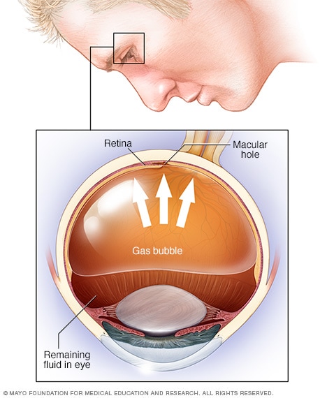
Pneumatic retinopexy
If your retina has detached, you'll need surgery to repair it, preferably within days of a diagnosis. The type of surgery your surgeon recommends will depend on several factors, including how severe the detachment is.- Injecting air or gas into your eye. In this procedure, called pneumatic retinopexy (RET-ih-no-pek-see), the surgeon injects a bubble of air or gas into the center part of the eye (the vitreous cavity). If positioned properly, the bubble pushes the area of the retina containing the hole or holes against the wall of the eye, stopping the flow of fluid into the space behind the retina. Your doctor also uses cryopexy during the procedure to repair the retinal break.Fluid that had collected under the retina is absorbed by itself, and the retina can then adhere to the wall of your eye. You may need to hold your head in a certain position for up to several days to keep the bubble in the proper position. The bubble eventually will reabsorb on its own.
- Indenting the surface of your eye. This procedure, called scleral (SKLAIR-ul) buckling, involves the surgeon sewing (suturing) a piece of silicone material to the white of your eye (sclera) over the affected area. This procedure indents the wall of the eye and relieves some of the force caused by the vitreous tugging on the retina.If you have several tears or holes or an extensive detachment, your surgeon may create a scleral buckle that encircles your entire eye like a belt. The buckle is placed in a way that doesn't block your vision, and it usually remains in place permanently.
- Draining and replacing the fluid in the eye. In this procedure, called vitrectomy (vih-TREK-tuh-me), the surgeon removes the vitreous along with any tissue that is tugging on the retina. Air, gas or silicone oil is then injected into the vitreous space to help flatten the retina.Eventually the air, gas or liquid will be absorbed, and the vitreous space will refill with body fluid. If silicone oil was used, it may be surgically removed months later.Vitrectomy may be combined with a scleral buckling procedure.
After surgery your vision may take several months to improve. You may need a second surgery for successful treatment. Some people never recover all of their lost vision.
Coping and support
Retinal detachment may cause you to lose vision. Depending on your degree of vision loss, your lifestyle might change significantly.
You may find the following ideas useful as you learn to live with impaired vision:
- Get glasses. Optimize the vision you have with glasses that are specifically tailored for your eyes. Request safety lenses to protect your better-seeing eye.
- Brighten your home. Have proper light in your home for reading and other activities.
- Make your home safer. Eliminate throw rugs and place colored tape on the edges of steps. Consider installing motion-activated lights.
- Enlist the help of others. Tell friends and family members about your vision problems so they can help you.
- Get help from technology. Digital talking books and computer screen readers can help with reading, and other new technology continues to advance.
- Check into transportation. Investigate vans and shuttles, volunteer driving networks, or ride shares available in your area for people with impaired vision.
- Talk to others with impaired vision. Take advantage of online networks, support groups and resources for people with impaired vision.
IClinix– Advanced Eye Care is driven by the quest to protect and prevent vision loss and blindness due to variety of eye disorders.
ReplyDeleteRetinal Detachment Treatment l Diabetic Retinopathy Treatment l Motiyabind operation in delhi l Retinal Detachment Surgery l Best Eye Hospital in Delhi l Retina Laser surgery